Whole Slide Scanner vs Microscope: Full Comparison

Compare whole slide scanners vs traditional microscopes in pathology. Learn key differences in workflow, image quality, scalability, LIS integration, and digital pathology adoption.
5 mins

Whole Slide Scanner vs Microscope: Full Comparison

TL;DR:

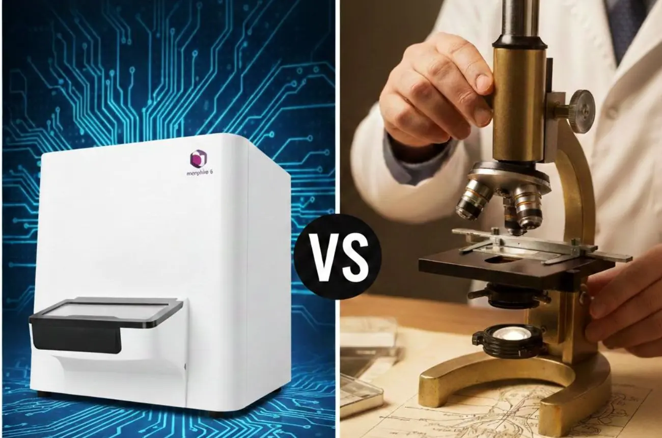
Traditional microscopes have long been the gold standard in pathology, but digital pathology scanners are rapidly redeining what's possible. This blog breaks down the key differences between whole slide scanners and conventional microscopes, covering workflow, image quality, scalability, and real-world applications, so you can make a confident, informed decision for your lab.

What You'll Learn

  • How whole slide scanners and traditional microscopes work
  • Key workflow and technical differences
  • Benefits and limitations of each approach
  • Real-world applications in histology and clinical pathology
  • What to look for when buying a digital pathology scanner

Defining the Two Technologies

A traditional microscope is an optical instrument that magnifies tissue samples on glass slides using lenses and light. Pathologists physically look through the eyepiece, adjusting magnification and focus manually. It's tactile, direct, and familiar, but inherently limited by geography, throughput, and the human eye.

A digital pathology scanner (also called a whole slide scanner or automated microscope slide scanner) converts glass slides into high-resolution digital images, known as Whole Slide Images (WSI), that can be viewed, analyzed, shared, and archived on any connected device. These scanners capture an entire slide at multiple magnifications with consistent, automated focus across every field of view.

Workflow: Manual vs. Automated

With a traditional microscope, a pathologist places a slide, selects magnification, adjusts the focus manually, and examines regions of interest one field at a time. This process is time-consuming and cannot scale easily.

A digital pathology scanner automates the entire capture process. Slides are loaded; either individually or in batches,  and the scanner produces a complete, navigable digital image in minutes. Once digitized, slides can be instantly shared with remote consultants, analyzed using AI-powered tools, or retrieved from a digital archive years later without degradation. The contrast in throughput alone is stark: high-end scanners can process hundreds of slides per day, while a single pathologist at a microscope is limited by physical endurance and time.

Technical Factors: Image Quality, Speed & Consistency

Both technologies are capable of producing high-quality images, but the conditions under which they do so differ significantly.

Traditional microscopes excel in real-time, dynamic observation. An experienced pathologist can rapidly scan a slide and intuitively navigate to areas of interest. However, image quality is dependent on the individual, the condition of the optics, and ambient lighting.

A digital pathology scanner delivers standardized, reproducible image quality regardless of the operator. Features like autofocus algorithms, multi-layer Z-stacking, and consistent illumination ensure every image is captured at the same quality benchmark. This consistency is critical in multi-site institutions, research collaborations, and teleconsultation workflows. A Slide Scanner for Histology settings, for example, must faithfully reproduce tissue architecture and staining patterns;  something modern scanners handle with precision at 20x and 40x equivalent magnifications.

Benefits vs. Limitations

Whole Slide Scanners — Benefits:

  • Remote access and teleconsultation capabilities
  • AI and image analysis integration
  • Permanent, lossless digital archives
  • Higher throughput and batch processing
  • Consistent image quality across operators and sites

Whole Slide Scanners — Limitations:

  • Higher upfront digital pathology scanner price compared to standard microscopes
  • Requires IT infrastructure and storage capacity
  • Learning curve for digitally-native workflows
  • Not all clinical environments are validated for primary diagnosis via WSI (though regulatory acceptance is growing rapidly)

Traditional Microscopes — Benefits:

  • Lower initial cost
  • No dependence on IT systems
  • Familiar to all pathologists
  • Effective for real-time, low-throughput environments

Traditional Microscopes — Limitations:

  • No remote access or sharing without additional imaging setups
  • Slides degrade over time; no permanent archive
  • Not scalable for high-volume labs
  • Cannot integrate with AI diagnostic tools

Applications: Where Each Technology Wins

Traditional microscopes remain effective in small clinics or settings with minimal digital infrastructure. However, for the majority of modern use cases, digital pathology scanners hold a clear advantage.

Histopathology and surgical pathologefit enormously from Whole slide imaging, enabling rapid frozen section review, second opinions from remote specialists, and streamlined tumor board presentations. Histology scanner systems are now standard in many academic medical centers for teaching and research. Clinical trials and pharmaceutical research increasingly mandate digital imaging for reproducibility. Cytology, hematology, and even dermatopathology are all being transformed by whole slide imaging.

Buying Guide: Key Considerations

When evaluating a digital pathology scanner for your institution, consider the following:

  • Scan speed and throughput : How many slides per hour does your workflow demand?
  • Resolution and magnification equivalents : Does it support 20x, 40x, or brightfield and fluorescence?
  • Software ecosystem : Does the scanner integrate with your LIS, PACS, or AI tools?
  • Digital pathology scanner price and TCO :  Evaluate the total cost of ownership, including storage, maintenance, and training
  • Regulatory compliance : Is the scanner FDA-cleared or CE-marked for primary diagnosis?
  • Vendor support and scalability :  Can the system grow with your lab?

The Future: AI, Automation & Digital-First Pathology

The trajectory is clear: pathology is going digital. Regulatory agencies like the FDA and the EU have increasingly validated WSI for primary diagnosis. AI-powered tools for tumor detection, grading, and biomarker quantification are rapidly maturing, and they all depend on high-quality digital slide data generated by digital pathology scanners.

Digital pathology companies at the forefront of this transition are not just building scanners, they're building ecosystems. The convergence of AI, cloud storage, and high-throughput scanning is redefining what a modern pathology lab looks like.

Why Morphle Labs

Transitioning to digital pathology isn’t just about replacing microscopes with scanners; it's about redesigning how slides move through your lab’s workflow. Morphle Labs’ whole slide scanners are built with this operational reality in mind.

Beyond high-resolution image capture, Morphle systems integrate directly into existing laboratory workflows through built-in case management and support for both uni-directional and bi-directional LIS integration. This allows labs to automate slide-to-case mapping using barcodes, QR codes, or even handwritten labels, reducing manual data entry and minimizing identification errors during case assembly.

For institutions managing multi-site reporting or remote sign-out, Morphle’s web-based viewing environment enables pathologists to access, review, and annotate whole slide images from any authorized device or location. Batch scanning capabilities further allow technical staff to digitize large volumes of slides with minimal operator intervention, freeing up skilled personnel for higher-value tasks.

By combining consistent 40x imaging with integrated workflow automation, Morphle’s digital pathology platform supports scalable deployment across clinical, research, and teaching environments. Whether you're building a digital archive, enabling teleconsultation, or preparing your lab for AI-assisted analysis, Morphle provides the infrastructure required for a practical transition to digital-first pathology.

.

Ready to Upgrade Your Lab?

The shift from traditional microscopes to digital pathology is no longer a question of if ; it's a question of when and with whom. If your lab is ready to scale, collaborate remotely, and leverage the power of AI-assisted diagnostics, a best-in-class whole slide scanner is your next step.

Explore Morphle Labs' digital pathology solutions →

Get in touch with the Morphle team today for a personalized demo, pricing information, and to find out how their scanners can integrate into your existing workflow. The future of pathology is digital, and it starts here.

Learn more about digital pathology and various usecases

Read More
Contact Us
Afghanistan (+93)
Albania (+355)
Algeria (+213)
American Samoa (+1-684)
Andorra (+376)
Angola (+244)
Anguilla (+1-264)
Antarctica (+672)
Antigua and Barbuda (+1-268)
Argentina (+54)
Armenia (+374)
Aruba (+297)
Australia (+61)
Austria (+43)
Azerbaijan (+994)
Bahamas (+1-242)
Bahrain (+973)
Bangladesh (+880)
Barbados (+1-246)
Belarus (+375)
Belgium (+32)
Belize (+501)
Benin (+229)
Bermuda (+1-441)
Bhutan (+975)
Bolivia (+591)
Bosnia and Herzegovina (+387)
Botswana (+267)
Brazil (+55)
British Indian Ocean Territory (+246)
Brunei (+673)
Bulgaria (+359)
Burkina Faso (+226)
Burundi (+257)
Cambodia (+855)
Cameroon (+237)
Canada (+1)
Cape Verde (+238)
Cayman Islands (+1-345)
Central African Republic (+236)
Chad (+235)
Chile (+56)
China (+86)
Christmas Island (+53)
Cocos Islands (+61)
Colombia (+57)
Comoros (+269)
Congo (DRC) (+243)
Congo (Republic) (+242)
Cook Islands (+682)
Costa Rica (+506)
Cote D'Ivoire (+225)
Croatia (+385)
Cuba (+53)
Cyprus (+357)
Czech Republic (+420)
Denmark (+45)
Djibouti (+253)
Dominica (+1-767)
Dominican Republic (+1-809 and +1-829  )
East Timor (+670)
Ecuador (+593 )
Egypt (+20)
El Salvador (+503)
Equatorial Guinea (+240)
Eritrea (+291)
Estonia (+372)
Ethiopia (+251)
Falkland Islands (+500)
Faroe Islands (+298)
Fiji (+679)
Finland (+358)
France (+33)
French Guiana (+594)
French Polynesia (+689)
Gabon (+241)
Gambia (+220)
Georgia (+995)
Germany (+49)
Ghana (+233)
Gibraltar (+350)
Greece (+30)
Greenland (+299)
Grenada (+1-473)
Guadeloupe (+590)
Guam (+1-671)
Guatemala (+502)
Guinea (+224)
Guinea-Bissau (+245)
Guyana (+592)
Haiti (+509)
Honduras (+504)
Hong Kong (+852)
Hungary (+36)
Iceland (+354)
India (+91)
Indonesia (+62)
Iran (+98)
Iraq (+964)
Ireland (+353)
Israel (+972)
Italy (+39)
Jamaica (+1-876)
Japan (+81)
Jordan (+962)
Kazakhstan (+7)
Kenya (+254)
Kiribati (+686)
Kuwait (+965)
Kyrgyzstan (+996)
Laos (+856)
Latvia (+371)
Lebanon (+961)
Lesotho (+266)
Liberia (+231)
Libya (+218)
Liechtenstein (+423)
Lithuania (+370)
Luxembourg (+352)
Macau (+853)
Macedonia (+389)
Madagascar (+261)
Malawi (+265)
Malaysia (+60)
Maldives (+960)
Mali (+223)
Malta (+356)
Marshall Islands (+692)
Martinique (+596)
Mauritania (+222)
Mauritius (+230)
Mayotte (+269)
Mexico (+52)
Micronesia (+691)
Moldova (+373)
Monaco (+377)
Mongolia (+976)
Montserrat (+1-664)
Morocco (+212)
Mozambique (+258)
Myanmar (+95)
Namibia (+264)
Nauru (+674)
Nepal (+977)
Netherlands (+31)
Netherlands Antilles (+599)
New Caledonia (+687)
New Zealand (+64)
Nicaragua (+505)
Niger (+227)
Nigeria (+234)
Niue (+683)
Norfolk Island (+672)
Northern Mariana Islands (+1-670)
North Korea (+850)
Norway (+47)
Oman (+968)
Pakistan (+92)
Palau (+680)
Palestinian State (+970)
Panama (+507)
Papua New Guinea (+675)
Paraguay (+595)
Peru (+51)
Philippines (+63)
Poland (+48)
Portugal (+351)
Puerto Rico (+1-787 or +1-939)
Qatar (+974 )
Reunion (+262)
Romania (+40)
Russia (+7)
Rwanda (+250)
Saint Helena (+290)
Saint Kitts and Nevis (+1-869)
Saint Lucia (+1-758)
Saint Pierre and Miquelon (+508)
Saint Vincent and the Grenadines (+1-784)
Samoa (+685)
San Marino (+378)
Sao Tome and Principe (+239)
Saudi Arabia (+966)
Senegal (+221)
Serbia (+381)
Seychelles (+248)
Sierra Leone (+232)
Singapore (+65)
Slovakia (+421)
Slovenia (+386)
Solomon Islands (+677)
Somalia (+252)
South Africa (+27)
South Korea (+82)
South Sudan (+211)
Spain (+34)
Sri Lanka (+94)
Sudan (+249)
Suriname (+597)
Svalbard and Jan Mayen Islands (+47)
Swaziland (+268)
Sweden (+46)
Switzerland (+41)
Syria (+963)
Taiwan (+886)
Tajikistan (+992)
Tanzania (+255)
Thailand (+66)
Togo (+228)
Tokelau (+690)
Tonga (+676)
Trinidad and Tobago (+1-868)
Tunisia (+216)
Turkey (+90)
Turkmenistan (+993)
Turks and Caicos Islands (+1-649)
Tuvalu (+688)
Uganda (+256)
Ukraine (+380)
United Arab Emirates (+971)
United Kingdom (+44)
United States (+1)
Uruguay (+598)
Uzbekistan (+998)
Vanuatu (+678)
Vatican City State (+418)
Venezuela (+58)
Vietnam (+84)
Virgin Islands, British (+1-284)
Virgin Islands, United States (+1-340)
Wallis and Futuna Islands (+681)
Western Sahara (+212)
Yemen (+967)
Zambia (+260)
Zimbabwe (+263)