Veterinary diagnostic labs are rapidly embracing digital pathology, and for good reason. A modern digital pathology scanner eliminates geographic barriers, accelerates diagnoses, and opens the door to AI-powered analysis. This guide walks you through everything you need to know before leaping.
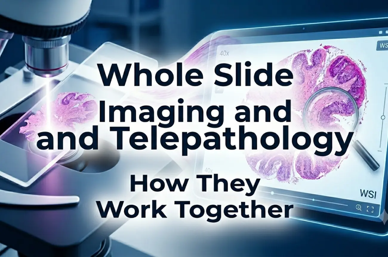
Veterinary digital pathology is the process of converting traditional glass histology slides into high-resolution digital images, called Whole Slide Images (WSIs), that can be stored, shared, and analyzed on a computer screen or cloud platform.
At its core, the process relies on a digital pathology scanner: a precision imaging instrument that captures an entire glass slide at magnifications equivalent to or exceeding a conventional optical microscope. The resulting file is navigable at any zoom level, just like a physical slide under a lens, but without the physical slide ever needing to leave the lab.
Once niche, digital pathology has rapidly evolved in veterinary medicine, with clinical pathology specimens now being routinely digitized alongside histology samples.
Understanding the end-to-end process helps set realistic expectations:
Sample collection → Preparation (fixation, embedding, microtomy, staining, mounting) → High-resolution scanning → Server upload → Remote review by the pathologist → Report generation
The digital pathology scanner sits at the critical hand-off point between the wet lab and the diagnostic workstation. Scan quality, throughput, and file format compatibility at this stage determine the success of everything downstream; from AI-assisted analysis to tele-consultation.
Before committing to any system, evaluate these parameters carefully:
Resolution and magnification — Most diagnostic applications require 20× or 40× equivalent scanning. Cytology specimens often demand higher magnification and Z-stack (multi-focal plane) capabilities, which not all scanners support.
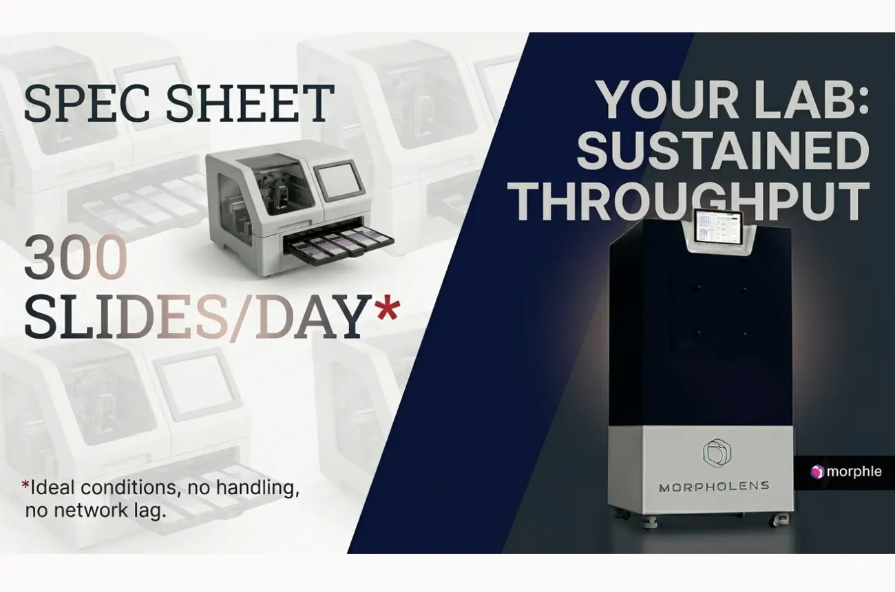
Throughput — High-volume diagnostic labs need automated batch scanning. An automated microscope slide scanner with multi-slide loading reduces technician time and standardizes image quality across runs.
File format and interoperability — Ensure your scanner outputs open or widely supported formats (SVS, NDPI, MRXS). Proprietary formats can create lock-in and complicate long-term data archiving.
Integration with LIS/LIMS — Seamless connection with your Laboratory Information System is non-negotiable for workflow efficiency and traceability.
Storage and viewing infrastructure — WSI files are large (1–4 GB per slide is common). Plan your server or cloud storage capacity before day one.
Benefits:
Limitations:
The use cases for digital pathology in veterinary settings span a wide spectrum:
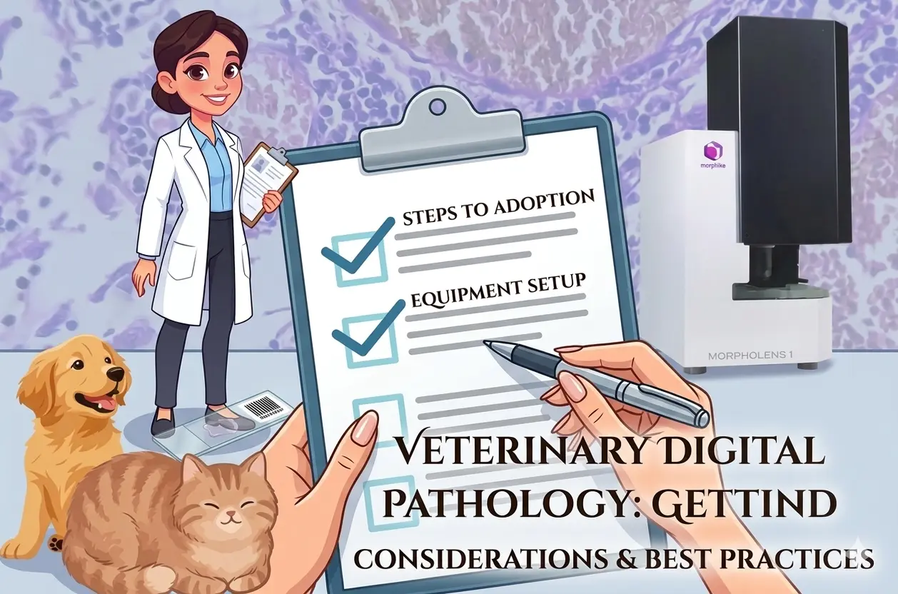
Use this checklist when evaluating vendors:
The field is moving fast. Key directions to watch:
AI-powered analysis — Machine learning models trained on veterinary WSI datasets are already demonstrating diagnostic accuracy comparable to experienced pathologists for specific tumor types. This will become routine.
Cloud-native pathology — Centralized scan-and-read models will allow regional diagnostic hubs to serve clinics across wide geographies without any glass slide logistics.
Multimodal integration — Combining digital morphology with genomics, proteomics, and clinical metadata will redefine prognostic scoring in veterinary oncology.
Point-of-care scanning — Smaller-footprint Histology Scanners are making it feasible to deploy scanning capability outside the central lab — at specialty clinics, research institutes, and even in the field.
Navigating the landscape of Digital Pathology Companies requires cutting through the noise. Morphle Labs is building precision scanning hardware with a clear focus on accessibility and performance — making whole slide imaging practical not just for large academic institutions but for mid-sized diagnostic labs and veterinary practices ready to modernize their workflows.
Morphle's digital pathology solutions are designed with interoperability and ease of integration in mind, so your team spends less time managing IT complexity and more time delivering diagnostic value.
The transition to digital pathology is no longer a question of if ; it's a question of when and with whom.
Whether you're scanning your first batch of histology slides or scaling up to a multi-site digital diagnostic network, getting the right digital pathology scanner from the right partner makes all the difference.
Explore Morphle Labs' veterinary pathology solutions today, and take the first step toward faster diagnoses, smarter workflows, and future-ready laboratory infrastructure.
Book a Demo with Morphle Labs →

.webp)
.webp)
.webp)
.webp)
.webp)
.webp)