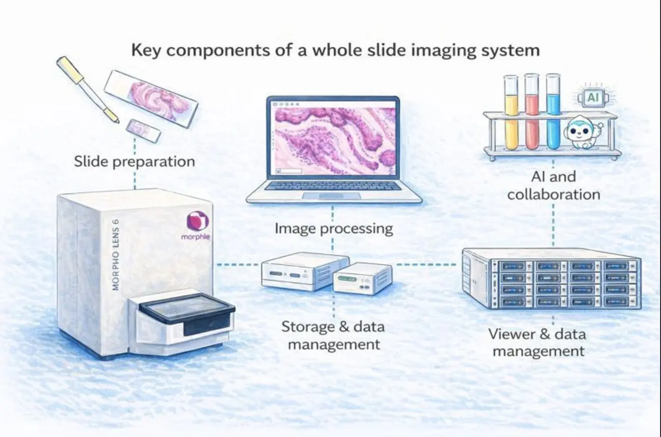
A whole slide imaging system (WSI) converts glass slides into high-resolution digital files that can be viewed, shared, analyzed, and archived. A complete WSI solution includes high-quality optical hardware, a reliable digital pathology scanner, efficient image processing, scalable storage, an intuitive WSI viewer, and workflow tools including AI and tele-pathology features. Understanding how these components work together helps laboratories choose a platform optimized for image quality, throughput, and diagnostic confidence.
Whole Slide Imaging (WSI)
Digitizing entire glass slides at high resolution for virtual microscopy.
Digital Pathology Scanner
Hardware that captures images of slides using precision optics, cameras, and automated mechanics.
WSI Viewer
Software that allows pathologists to pan, zoom, annotate, collaborate, and analyze slides; ideally with microscope-like responsiveness.
Telepathology
Reviewing and diagnosing slides remotely via secure streaming or shared digital files.
DICOM Pathology
Standards that enable interoperability with LIS/PACS systems and long-term archival of digital slides.
AI in Histopathology
Algorithms designed to assist with tissue detection, quantification, quality control, and decision support.
Whole slide imaging is more than scanning; it represents a full digital workflow transformation:
Each step affects diagnostic quality, turnaround time, and long-term scalability. Let’s break them down.
Optics determine whether critical diagnostic structures are visible; from nuclear morphology to IHC marker expression.
Key optical characteristics include:
High-quality optics reduce rescans, minimize uncertainty, and improve diagnostic confidence. Modern digital pathology scanners, including platforms such as Morphle’s, prioritize optical stability to ensure consistent performance across routine and high-volume workflows.
Once light passes through the optics, cameras and sensors convert it into digital data.
Important specifications:
A well-matched optics-camera combination supports both throughput and image fidelity; especially important for labs processing large daily volumes.
Whole slide imaging requires capturing thousands of microscopic fields of view with micron-level accuracy.
Critical mechanical components include:
As case volumes grow, scanner reliability and uptime become as important as raw scan speed. Vendors like Morphle design scanners with workflow continuity in mind, ensuring consistent performance during extended scanning runs.
Image processing software merges thousands of image tiles into a seamless whole slide image.
Core processing tasks include:
Smooth panning and instant tile loading begin at this stage. Efficient processing directly impacts the responsiveness of the WSI viewer downstream.
A single whole slide image can range from 0.5 to 2.5 GB. At scale, storage planning becomes critical.
Common deployment models:
File format considerations:
Modern WSI platforms; including Morphle’s; support flexible deployment options so labs can scale storage without locking themselves into a single architecture.
The viewer is where pathologists spend most of their time, and adoption depends heavily on performance.
Key features to look for:
Even the most advanced scanner hardware can fail adoption if the viewer feels laggy or unintuitive. Many modern systems, including Morphle’s digital pathology solutions, emphasize low-latency streaming and responsive interfaces to mimic the experience of traditional microscopy.
Digital pathology succeeds when it integrates seamlessly into existing laboratory processes.
Key workflow capabilities:
Telepathology enables faster access to subspecialty expertise, improving turnaround time and diagnostic safety; especially for distributed or resource-limited labs.
AI does not replace pathologists—it supports them.
Common applications include:
When integrated thoughtfully into the workflow, AI improves consistency and enables scalable analysis without disrupting diagnostic judgment.
When evaluating a whole slide imaging system, consider:
Throughput & scalability
Can the system handle today’s volumes and future growth?
Viewer responsiveness
Does it provide a microscope-like experience on standard devices?
Deployment flexibility
Does it support on-prem, cloud, or hybrid architectures?
Integration readiness
Will it fit smoothly into existing LIS/PACS workflows?
Total cost of ownership
Consider storage, service contracts, AI modules, maintenance, and uptime guarantees. A system that appears cheaper upfront may cost more operationally over time.
Digital pathology is a transformation; not just a purchase.
Morphle’s digital pathology platform is designed to align with the core components of a modern whole slide imaging system; focusing on reliability, usability, and scalability rather than isolated features.
Morphle scanners combine high-quality optics, precise mechanics, and stable illumination to deliver consistent image quality across routine and high-volume workflows. The image processing and viewing pipeline is optimized for smooth stitching, fast pan-and-zoom performance, and a microscope-like user experience that supports confident digital sign-out.
To accommodate diverse IT environments, Morphle supports on-premises, cloud, and hybrid deployment models, allowing laboratories to scale storage while maintaining control over data and compliance. Built-in workflow integration, secure remote access, and AI-ready architecture further enable telepathology, collaboration, and future adoption of computational pathology tools.
A successful whole slide imaging system unifies:
Together, these components enable pathologists to work faster, collaborate more effectively, and unlock new diagnostic insights through virtual microscopy and telepathology.
Explore how Morphle supports laboratories with flexible deployment options and a 30-day risk-free evaluation.

.webp)
.webp)
.webp)
.webp)
.webp)
.webp)