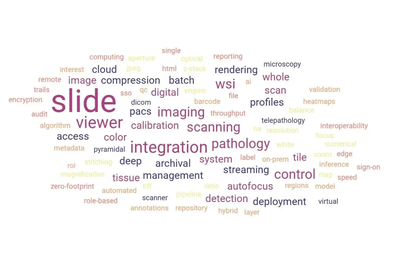
Digital pathology is filled with technical terminology that can feel overwhelming, WSI, DICOM pathology, tele-pathology, AI pipelines, rendering engines, compression types, and more. This guide breaks down the most essential terms in simple, accessible language so pathologists, lab managers, and researchers can confidently evaluate a whole slide imaging system and understand how these components shape workflow, accuracy, and scalability.
A combination of hardware and software that converts glass slides into high-resolution digital images. It includes the scanner, the optics, the robotics, calibration tools, image processing engines, and the viewer. This is the backbone of modern digital pathology.
The device that physically scans histology slides. It captures a complete, high-fidelity image of the entire tissue section—stitched, focused, color-corrected, and ready for diagnosis.
A software viewer that lets pathologists navigate the digital slide as smoothly as a microscope, with pan, zoom, annotations, measurement tools, and side-by-side comparison. Speed and responsiveness are critical because delays interrupt diagnostic flow.
The practice of examining digital slides instead of glass slides. Virtual microscopy replicates the microscope experience while adding digital advantages like instant sharing, overlays, and AI assistance.
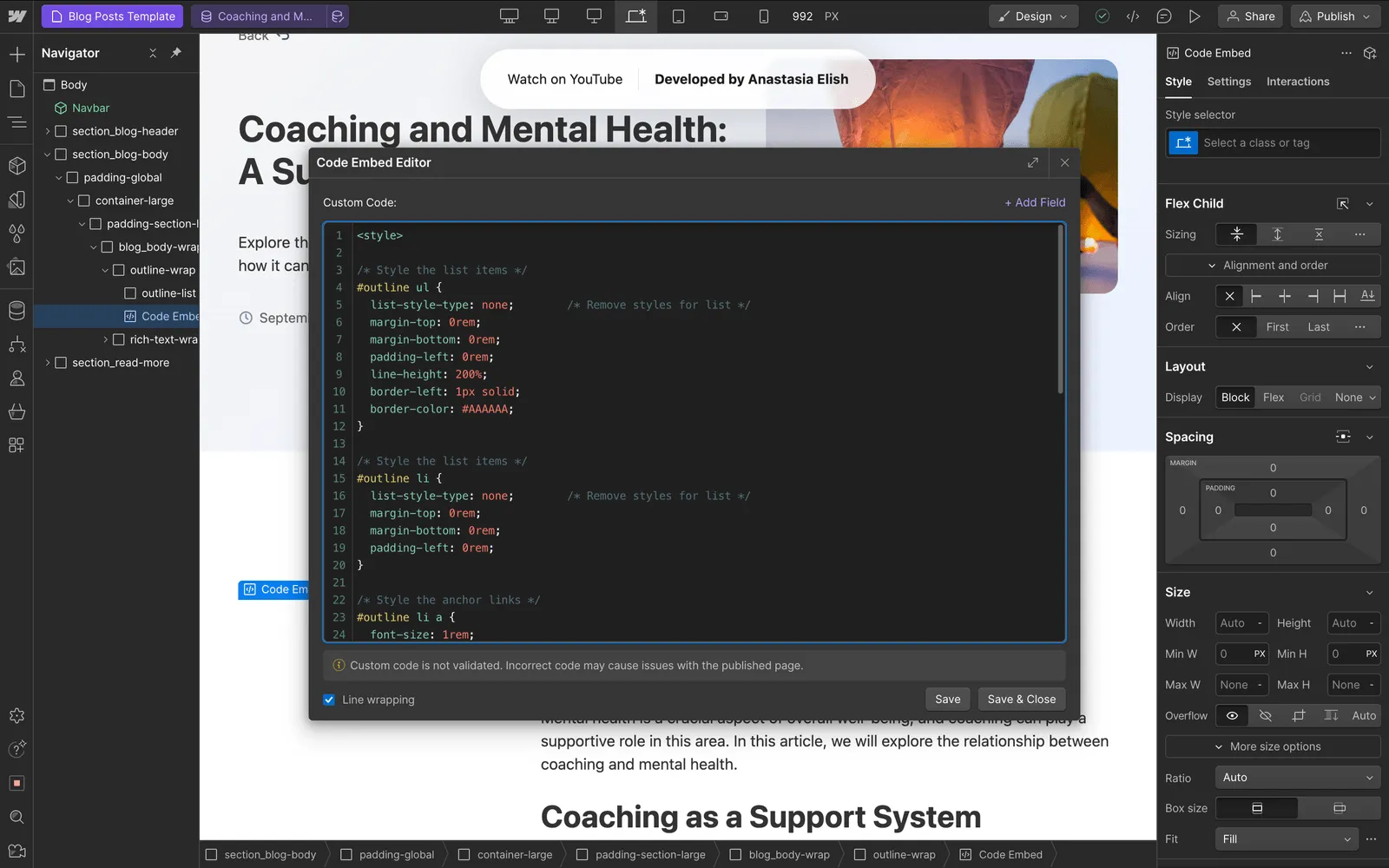
The remote review or consultation of digital slides. Morphle scanners have been used across continents to enable real-time collaboration—an experience described in our story here.
The standard for managing, storing, and transmitting medical images. DICOM ensures compatibility across systems and long-term archival integrity—a key requirement for enterprise adoption.
Machine learning tools that assist diagnosis by detecting patterns, quantifying biomarkers, scoring features, or aiding triage. AI models depend heavily on image consistency and metadata quality.
Digital pathology is more than scanning; it is a complete pipeline.
- A slide enters the scanner, where precision optics capture multi-focal layers.
- The system automatically selects the best focus, stitches tiles, balances colors, and generates a clean digital file.
- The WSI viewer then streams the image without lag, whether the user is on a workstation or a mobile device.
- Tele-pathology tools make it possible for experts anywhere in the world to read the case instantly.
- Finally, archival systems store the image securely, often in DICOM format, ready for retrieval months or years later.
Every term in digital pathology maps to one phase of this journey; capture, process, view, share, analyze, archive.
Higher magnification does not guarantee diagnostic clarity. Optical design, NA, sensor quality, and focus algorithms matter more. Consistent resolution across the entire tissue section is what enables trustworthy analysis.
Modern scanners use predictive auto-focus, multi-plane scanning, or hybrid strategies combining machine learning and optical mapping. This ensures crisp cellular detail even in uneven tissue.
Most images are built from thousands of tiles. Seamless stitching, color calibration, shading correction, and noise reduction determine how “microscope-like” the final image feels.
WSI files are huge. JPEG2000, pyramidal TIFF, and proprietary formats all have trade-offs between quality, loading time, and storage efficiency. These choices impact downstream AI use.
A WSI viewer should not lag, buffer, or blur excessively during zoom transitions. Rendering pipelines, caching, and network optimization define the real-world experience.
Authentication, encrypted transfers, role-based access, and full audit trails ensure safe tele-pathology and protect patient data.
Digital pathology brings speed, accessibility, and scalability, but it also introduces challenges.
Understanding the terminology helps teams select systems that minimize these limitations.
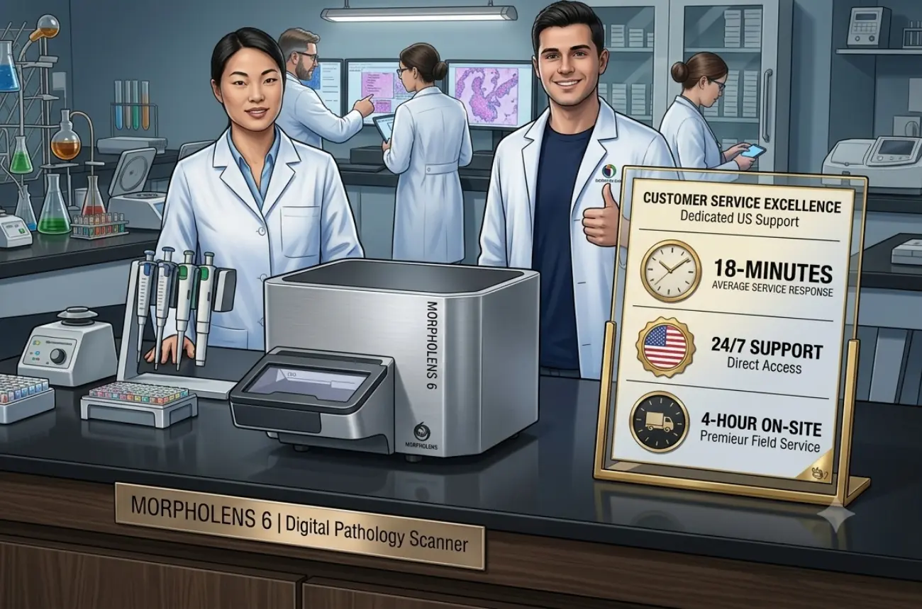
Digital pathology must align with standards:
Compliance terminology ensures your investment remains future-proof as digital pathology becomes mainstream.
Digital workflows amplify value across specialties:
Tele-pathology especially transforms turnaround time, enabling senior pathologists to support satellite centers instantly.
When considering a whole slide imaging scanner, pay attention to terms such as:
For a deeper overview of choosing the right scanner : Explore options
Digital pathology is evolving quickly. New terminology will soon become routine:
These innovations will make tele-pathology even more seamless; an area where Morphle excels, especially in environments where consistency, reliability, and ease of use matter most.
Understanding the terminology behind digital pathology empowers labs, hospital health system and lab operations to adopt technology confidently.
A whole slide imaging system is not one device; it’s an ecosystem of scanning precision, viewer performance, secure sharing, and long-term interoperability.
Morphle makes this ecosystem simple, intuitive, and tele-pathology-ready.
Contact our Specialists for a right-sized WSI path or request a demo.

.webp)
.webp)
.webp)
.webp)
.webp)
.webp)