Digital pathology scanner accuracy depends on multiple interconnected factors: color reproducibility, spatial resolution, focusing precision, scanning methodology, image quality, and the expertise of operating personnel. Understanding these variables is essential for laboratories selecting and validating whole slide imaging systems that meet diagnostic standards while maintaining consistency across different platforms and workflows.
The transition from traditional microscopy to digital pathology represents a fundamental shift in how pathologists make diagnoses. Unlike conventional microscopy where pathologists intuitively compensate for image variations, a digital pathology scanner must maintain stable, reproducible image quality at levels required for clinical diagnosis. This makes accuracy not just desirable; it's mandatory for patient safety.
Scanner accuracy encompasses several critical performance characteristics. Color reproducibility ensures that tissue stains appear consistent across different scans and viewing sessions. Spatial resolution determines how effectively the system distinguishes between adjacent cellular structures. Focusing accuracy affects whether nuclear and cytoplasmic details remain sharp throughout the digitized slide. Together, these factors determine whether a whole slide scanner can reliably replicate what pathologists see under traditional microscopes.
The objective lens serves as the cornerstone of any digital pathology scanner. High-quality optics from established manufacturers deliver superior resolution and color fidelity. The imaging sensor's ability to capture data; whether through continuous motion or stop-and-capture methods; directly influences both speed and precision. Motion systems must achieve sub-micron precision to prevent image artifacts that compromise diagnostic quality.
Pathological specimens rely on staining to enhance optical visibility of tissue structures, making color reproducibility one of the most critical quality factors. Variations in color representation can lead pathologists to misinterpret cellular characteristics, potentially affecting diagnosis. Proper white balance calibration and spectral measurement protocols ensure that tissue evaluation points maintain true color values across different scanning sessions and display monitors.
Traditional microscopy allows pathologists to focus through the tissue's z-plane, examining nuclear details at multiple depths within a 4-5 micron section. Most digital scans capture a single focal plane, which can limit evaluation of certain specimens. Advanced automated microscope slide scanners address this through dense focus mapping and multi-layer z-stack acquisition, capturing 7-11 focal planes to preserve diagnostic information that might otherwise be lost.
After capturing individual fields, scanners must seamlessly stitch images together into a continuous virtual slide. The stitching algorithm's precision affects whether tissue boundaries remain intact and cellular patterns stay uninterrupted. File compression methods must balance storage efficiency against image quality; excessive compression introduces artifacts, while uncompressed files consume prohibitive storage space.
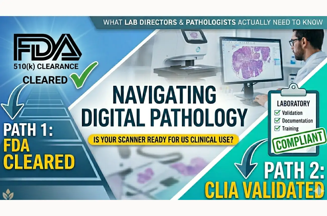
Regulatory bodies and professional organizations establish validation standards that laboratories must meet before deploying digital pathology for primary diagnosis. Studies demonstrate weighted mean concordance rates of 95.2% between whole slide imaging and optical microscopy, though concordance varies by specimen type and scanner platform.
Comparative assessments show that major diagnostic discordances occur at rates between 2.08% for optical microscopy and 2.4% for digital systems, with statistically insignificant differences between platforms when properly validated. Cytology specimens present particular challenges, requiring enhanced scanning protocols and additional pathologist training to achieve reliable accuracy.
Scanner accuracy extends beyond hardware specifications. Pathologists initially spend more time with digital slides due to unfamiliarity, learning curves, screen loading times, and digital navigation requirements. However, diagnostic time decreases substantially with practice and training.
Quality control processes must monitor both pre-imaging factors (slide preparation, tissue sectioning) and imaging factors (scanner calibration, focus accuracy) daily. Rescan rates typically range from 1-2% due to barcode detection failures, tissue detection issues, or image quality concerns. Laboratories must establish protocols to identify and address these technical failures promptly.
When accuracy standards are met, digital pathology delivers substantial advantages. Remote consultation becomes seamless, enabling subspecialty expertise regardless of geographic location. Archived digital slides remain accessible indefinitely without the stain degradation that affects glass slides. Multiple section levels and special stains can be viewed simultaneously, helping pathologists track focal abnormalities across different preparations.
AI integration represents perhaps the most transformative benefit. Algorithms can identify, quantify, and characterize diagnostic entities with consistency that reduces inter-observer variability. These tools surface subtle findings that might otherwise be missed, potentially improving detection rates for treatment-relevant disease.
When evaluating pathology slide scanners, laboratories should prioritize several key specifications. Resolution should reach 0.22 microns per pixel at 40x magnification. Scan times for standard specimens should complete within 2-3 minutes while maintaining image quality. The system must handle various slide types, including those with coverslip irregularities, uneven tissue sections, or non-standard dimensions.
Storage architecture matters significantly. Efficient compression algorithms can reduce file sizes to 0.1-0.5 gigabytes per whole slide image without sacrificing diagnostic information. Network caching and intelligent compression enable lag-free navigation even with limited bandwidth.
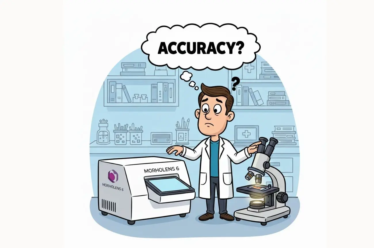
For laboratories seeking an accessible entry point into digital pathology, solutions like Morphle Labs offer compelling value propositions. Their platforms deliver image quality and scanning speed comparable to systems costing significantly more, with resolution reaching 0.22 microns per pixel and scan times averaging 90-250 seconds depending on focus density. The compact file format reduces storage costs by approximately 75% compared to conventional whole slide scanning formats. Multiple scanner configurations accommodate different laboratory volumes; from single-slide research units to 240-slide high-throughput systems; while maintaining consistent imaging standards.
The field continues evolving rapidly. Next-generation sensors promise faster capture rates without compromising precision. Enhanced autofocus algorithms reduce scanning time while improving focus accuracy across challenging specimens. Integration with laboratory information systems streamlines workflows and embeds patient data directly into digital pathology viewers.
Real-time quality assurance powered by edge AI can detect and correct image quality issues during acquisition, eliminating many rescans. Volumetric imaging capabilities will become standard, preserving the z-plane information that pathologists value from traditional microscopy. These advancements will further narrow any remaining accuracy gaps between digital and optical methods.
Accuracy isn't a single specification; it's the product of optical excellence, intelligent software, rigorous validation, and proper implementation. Laboratories evaluating whole slide imaging systems should conduct comprehensive assessments across multiple specimen types, involve stakeholders in hands-on testing, and ensure vendors provide robust training and support.
Ready to explore how digital pathology can transform your laboratory's diagnostic capabilities? Contact Morphle Labs for a demonstration of their whole slide scanning platforms. Their team provides validation support, workflow integration guidance, and ongoing calibration assistance to ensure your digital pathology implementation delivers the accuracy your patients deserve. Visit morphlelabs.com or reach out to sales@morphlelabs.com to schedule your consultation today.

.webp)
.webp)
.webp)
.webp)
.webp)
.webp)