Automated slide scanners are transforming histology by digitizing glass slides into high-resolution whole-slide images, enabling faster diagnosis, remote reporting, and AI-driven insights. As digital pathology adoption accelerates globally, a Digital Pathology slide scanner is becoming a foundational tool for modern labs.
For a deeper primer on digital pathology, explore: Knowledge Hub
A device that automatically loads, scans, and converts glass histology slides into digital images.
A scanner designed specifically for pathology workflows, optimized for color fidelity, resolution, and diagnostic accuracy.
The technology that captures the entire tissue section at high resolution, allowing pathologists to view it digitally.
A system tailored for routine histopathology slides, often used in clinical labs and high-volume workflows.
A scanner integrated with pathology systems, enabling seamless reporting, case tracking, and telepathology.
Automated slide scanners streamline the histology workflow by reducing manual handling and variability. After tissue processing and staining, slides are loaded into the scanner—either singly or in batches. The system detects tissue, sets focus points, optimizes illumination, and captures thousands of high-resolution fields that are stitched into a single whole-slide image.
These digital images are then uploaded to a secure viewer or LIS-integrated system. Pathologists can access the slides instantly, whether they are in the same facility or working remotely. Automated scanning eliminates bottlenecks caused by slide transport, microscope availability, and geographic constraints.
This shift is especially valuable for multi-site hospitals, high-volume labs, and centers exploring telepathology or AI-assisted workflows.
Automated slide scanners differ widely in performance. Understanding the technical concepts helps labs make informed decisions.
High-quality objectives and sensors determine image clarity. A 20X or 40X objective is common, but optical quality matters more than magnification alone.
Uneven tissue thickness demands advanced autofocus systems, including predictive focus maps and multi-layer scanning.
Accurate color calibration ensures stains appear consistent across devices and sessions—critical for diagnosis.
Whole-slide images can reach gigabytes in size. Efficient compression and streaming ensure smooth viewing without compromising detail.
Speed varies by tissue type, resolution, and scanner design. In high-volume labs, throughput is a primary selection factor.
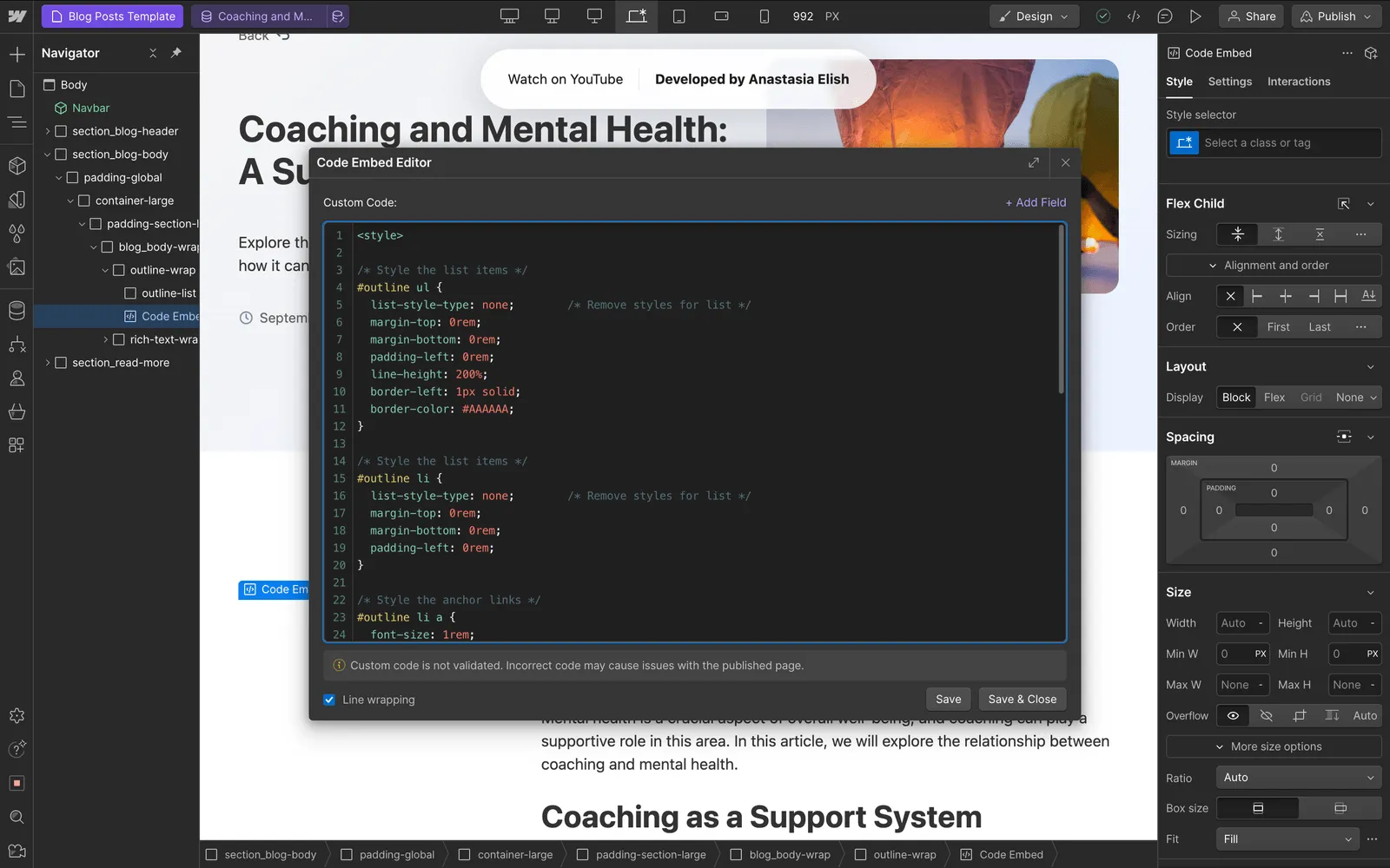
A responsive, microscope-like pan-and-zoom experience is essential. Morphle scanners excel in this area, offering fluid navigation on any device with on-prem, cloud, or hybrid deployment.
Automated slide scanners enhance consistency, reduce manual errors, and increase lab throughput. They enable remote consultations, support teaching and tumor boards, and prepare labs for AI-assisted diagnostics. Digital slides do not degrade over time, eliminating risk of breakage, fading, or misplacement. Automated workflows reduce technician handling and ensure superior traceability.
Barriers include initial investment, storage requirements, and IT infrastructure upgrades. Some labs may need cultural or workflow adaptation during the transition from microscopes to digital viewers. Scanning artifacts can occur if slides are improperly prepared. Despite these challenges, long-term efficiency gains outweigh most limitations.
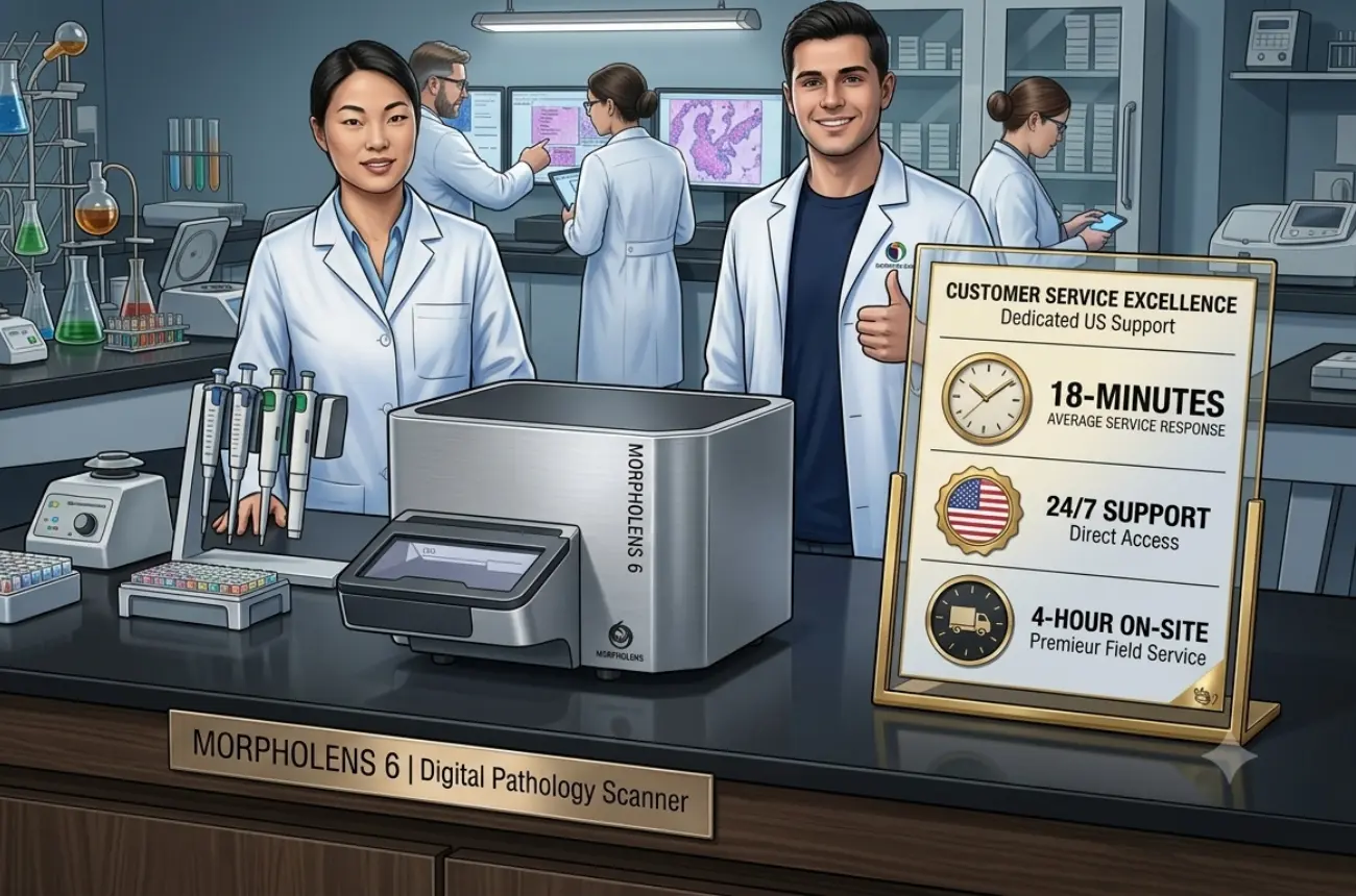
Digital pathology must align with regulatory frameworks such as HIPAA or GDPR for data protection, as well as CAP guidelines and internal quality policies. Consistent calibration, documented validation, and audit logs are essential.
DICOM pathology is increasingly important for interoperability, archival compliance, and future AI compatibility. Selecting scanners that support secure workflows, encryption, and integration with LIS/PACS systems is critical for long-term sustainability.
Automated slide scanners support a wide range of use cases:
They are especially useful where case volumes are high or where pathologists need rapid access to subspecialty opinions.
When selecting a scanner, consider these factors:
For labs evaluating mid-volume automated scanning, explore : Robust 6-slide scanner
The future of histology is fully digital. Innovations include AI-native scanners, real-time QC detection, automated region-of-interest scanning, and cloud-first WSI platforms. Zero-footprint viewers, predictive scanning algorithms, and AI-enhanced focusing will further reduce turnaround times.
As these technologies evolve, scanners will become faster, more accurate, and easier to integrate—driving widespread adoption even in smaller regional labs.
Morphle scanners make automated histology scanning practical and scalable. With robust optics, precise focus control, and a microscope-like viewing experience, Morphle delivers consistent whole-slide image quality across routine and high-volume workflows. Flexible on-prem, cloud, and hybrid deployments allow labs to adopt digital pathology without disrupting existing infrastructure. Designed for reliability and throughput, Morphle helps histology labs transition smoothly to digital workflows while staying ready for tele-pathology and AI adoption.
Automated slide scanners are reshaping histology, enabling faster workflows, greater collaboration, and AI-ready datasets. The best digital Pathology slide scanner for your lab is no longer a luxury—it is becoming essential infrastructure for modern pathology. With microscope-like navigation, flexible deployment options, and a responsive viewing experience, Morphle scanners make digital workflows accessible and scalable for any lab.
See automated scanning in action—book a live demonstration.

.webp)
.webp)
.webp)
.webp)
.webp)
.webp)